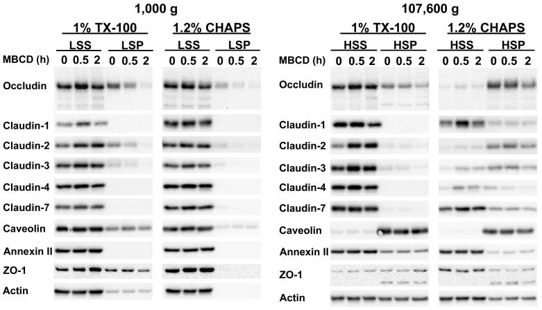
Figure 1.
Western blot analysis of MDCK II cell monolayers incubated with 10 mM MBCD for 0, 0.5 or 2 h. Monolayers were lysed either in TX-100 or CHAPS at 4°C and centrifuged at 1000 g for 30 min. Low speed pellets (LSP) and aliquots of the low speed supernatants (LSS) were retrieved. The remaining low speed supernatants were centrifuged at 4°C for 18 h at 107,600 g. High-speed supernatants (HSS) and high-speed pellets (HSP) were harvested. All supernatant and pellet fractions were processed for Western blotting and probed for occludin, claudin-1, -2, -3, -4, -7, caveolin-1, annexin-2, ZO-1 and actin. The data shown are representative of those obtained from a minimum of two independent experiments using TX-100 and CHAPS.

