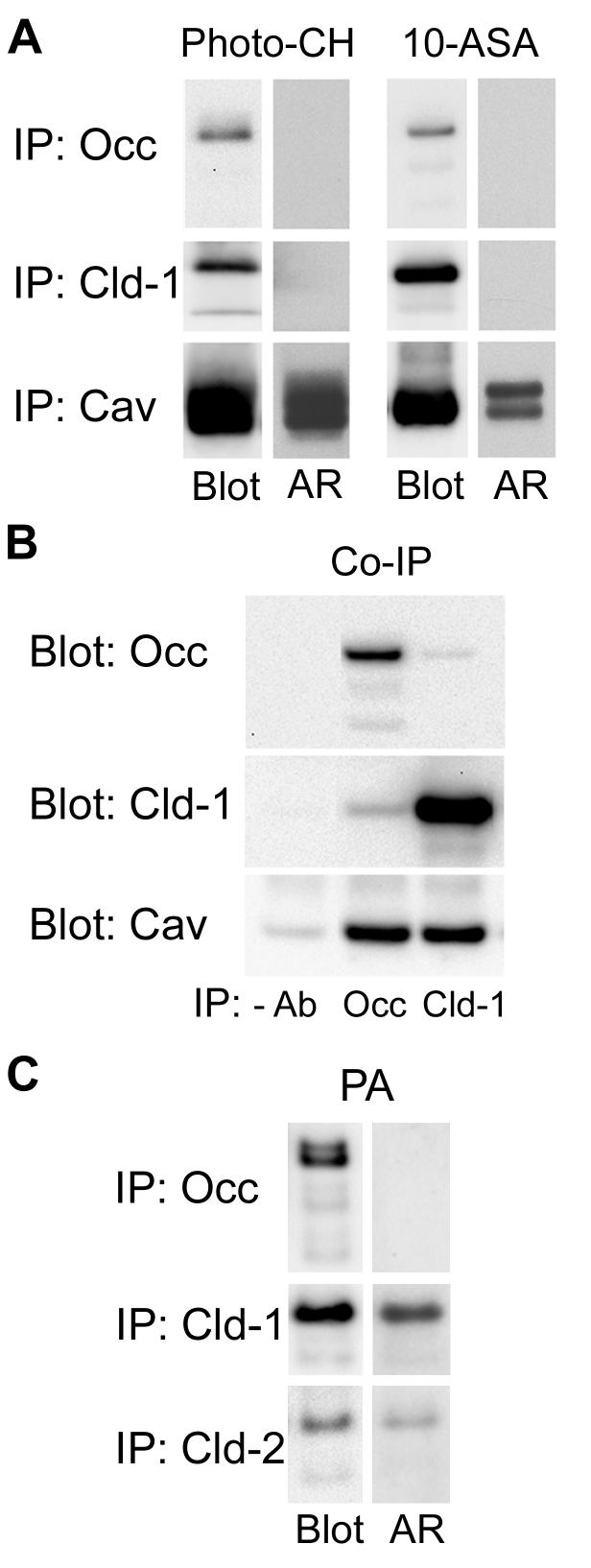
Figure 4.
A. MDCK II cell monolayers were incubated with either [3H]-photo-CH or [3H]-choline and 10-ASA as described in Materials and Methods. Following photo-activation, occludin, claudin-1 and caveolin-1 were sequentially immunoprecipitated (IP) in RIPA buffer. Dried PVDF membranes were subjected to autoradiography (AR) and probed for the respective immunoprecipitated protein (Blot). Whereas [3H]-photo-CH and [3H]-photo-PC prominently labeled caveolin-1, neither occludin nor claudin-1 were labeled. B. Occludin or claudin-1 was immunoprecipitated from lysates in 1% CHAPS, 0.05% SDS and the blots were probed with anti-occludin, anti-claudin-1 or anti-caveolin-1. Caveolin-1 is co-immunoprecipitated by both anti-occludin and anti-claudin-1. C. MDCK II cell monolayers were metabolically labeled with [3H]-palmitic acid (PA) as described in Materials and Methods. Occludin, claudin-1 and claudin-2 were sequentially immunoprecipitated. Dried PVDF membranes were subjected to autoradiography (AR) and probed for the respective immunoprecipitated protein (Blot). [3H]-palmitate labels claudin-1 and -2 but not occludin.

