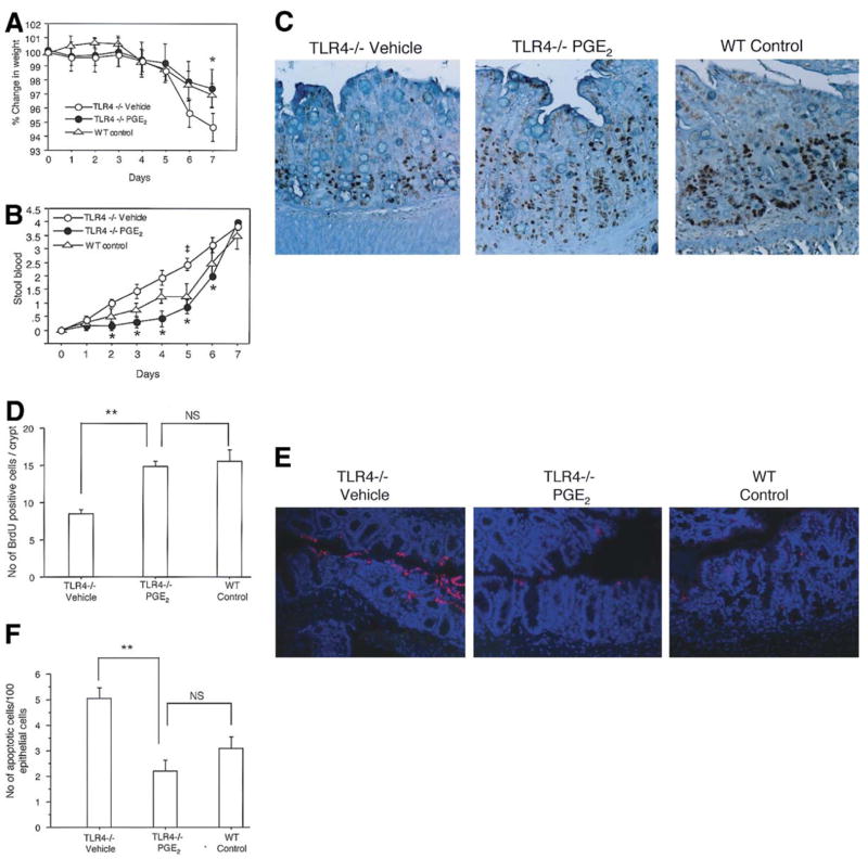
Figure 4. PGE2 supplementation improves signs of colitis and restores epithelial healing after DSS-induced injury in TLR4 −/− mice.
A. Weight change was examined daily during the 7 days of DSS treatment. Vehicle (PBS) treated TLR4−/− mice have significantly more weight loss than WT control mice (*P < 0.05). PGE2 treated TLR4−/− mice and have significantly less weight loss than vehicle treated TLR4−/− mice and are comparable to WT mice. The data represents the average (± SEM) of three independent experiments with a total of 19 mice (TLR4−/− Vehicle (n=6), TLR4−/− PGE2 (n=7) and WT controls (n=6)).
B. PGE2 treated TLR4−/− mice had a significant reduction in bleeding on days 2 through 6 compared with vehicle (PBS) treated TLR4−/− mice (*p<0.05). Stool blood was calculated as follows: 0=no blood, 1=trace occult blood positive, 2= strongly occult blood positive, and 4=bloody diarrhea. Standard error is shown.
C. BrdU labeling of intestinal epithelial cells was performed for vehicle treated TLR4−/−, PGE2 treated TLR4−/− and WT controls at the end of 7 days of DSS treatment. (Original magnification 200X)
D. BrdU positive cells were counted per HPF in 3 crypts of each colon segment (9 areas / mouse). Bars show mean ± SEM of proliferating cells / crypts (n=5 in each group). There is a significant increase in BrdU positive cells in PGE2 -treated TLR4−/− mice compared with vehicle treated TLR4−/− mice (**P < 0.001). PGE2-treated TLR4−/− mice are not significantly different than WT mice.
E. Apoptotic cells in the crypt epithelial cells were determined by TUNEL assay. Representative sections were taken from vehicle treated TLR4−/−, PGE2-treated TLR4−/− mice and WT controls as indicated. Red staining of nuclei indicates apoptotic cells. Sections were counterstained with DAPI (blue) to identify the nuclei of epithelial cells. PGE2 treated TLR4−/− mice showed a marked decrease of apoptotic cells compared with vehicle treated TLR−/− mice. The frequency of TUNEL positive epithelial cells in PGE2 treated TLR4−/− mice was similar to WT controls (original magnification 200X).
F. Number of apoptotic cells was counted in 300 total epithelial cells in triplicate in every 3 areas for each colon segment (n=5 in each group). Bars show mean ± SEM of the number of apoptotic cells per 100 total nuclei in the epithelial cells counted. There is a significant decrease of apoptotic cells in PGE2 treated TLR4−/− mice compared with vehicle treated TLR4−/− mice (**P < 0.001).

