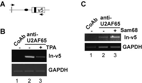
Figure 5. Sam68 and pre-mRNA occupancy by U2AF.
(A) Schematic drawing of the examined CD44 minigene pre-mRNA region (CD44 exon v5, open box; upstream intron, line; Sam68 binding sites, black oval and box, respectively; arrows, PCR primer). (B) RNP immunoprecipitations with an anti-U2AF65 antibody or a control antibody (CoAb) from lysates of LB17 lymphoma cells that were transfected with the pETv5 minigene construct containing CD44 exon v5. Cells were left either untreated (−) or treated with phorbol ester (TPA). TPA treatment and RNP immunoprecipitations were performed as described in legend to Figure 4. (C) RNP immunoprecipitations of U2AF65 from LB17 lymphoma cells co-transfected with the pETv5 minigene construct and either a Sam68 expression plasmid (+) or the empty expression vector as a control (−). Bands correspond to the expected sizes of 600 bp (In-v5) and 278 bp (GAPDH). All PCR amplifications were in linear phase as verified with different amounts of cDNA.

