
Figure 1.
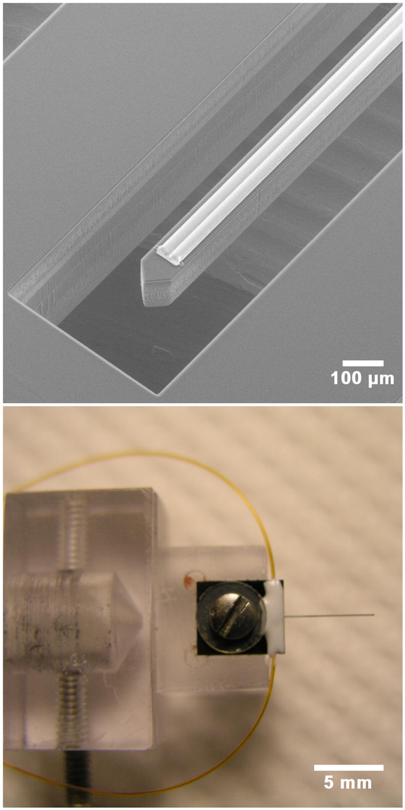
Upper: An electron micrograph of the probe showing the silicon shank and two parallel channels on top of the shank. The outlets of the channels are 0.5 mm from the shank tip. Lower: The probe is shown fixed to a custom holder by a screw and washer. Two small-bore polyimide tubes can be seen outside the holder. Each tube delivers fluid to a channel on the probe.