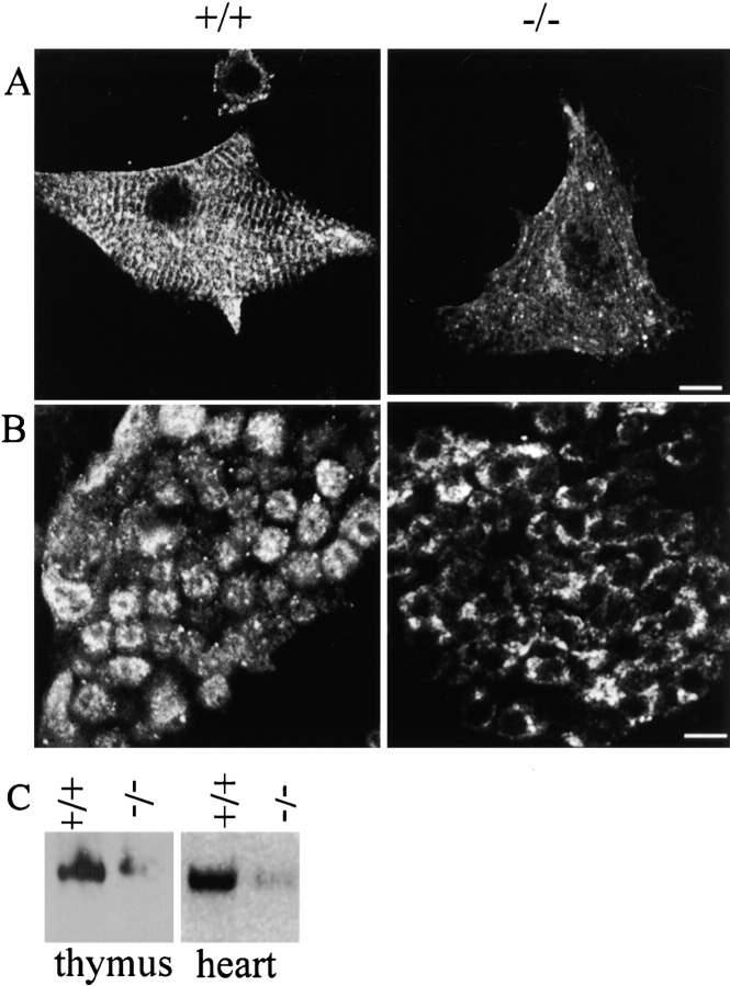
Figure 10.
Abnormal localization and reduced accumulation of IP3 receptors in cardiomyocytes and thymus of ankyrin-B (−/−) mice. Cardiomyocytes (A) and sections of thymus (B) of ankyrin-B (+/+) (left) and ankyrin-B (−/−) mice are labeled with antibody against IP3 receptor (types 1, 2, and 3) and visualized by immunofluorescence. Levels of label were computationally enhanced about twofold in ankyrin-B (−/−) examples to reveal localization. C shows immunoblots of equivalent amounts of ankyrin-B (+/+) and (−/−) thymus (left) and heart (right) with antibody against the IP3 receptor. Bars: (A) 10 μm; (B) 5 μm.

