
Figure 11.
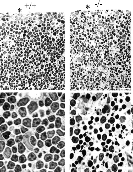
Toluidine blue–stained plastic sections of day 1 thymus from ankyrin-B (+/+) and (−/−) mice. Top panels are located at the cortex. An asterisk marks the margin of the thymus. Higher magnification images are shown at the bottom. Note that most of the nuclei of ankyrin-B (−/−) lymphocytes are condensed and pyknotic. Bars: (top) 25 μm; (bottom) 10 μm.