
Figure 6.
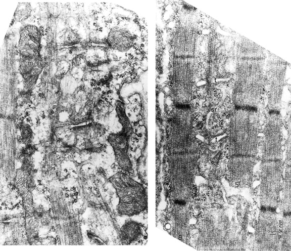
T-tubule and SR compartments and junctions between them are present in ankyrin-B (−/−) skeletal muscle. Electron micrographs (see Materials and Methods) of quadriceps muscle comparing the organization of the T-tubules and sarcoplasmic reticulum in littermates of normal (A) and ankyrin (−/−) (B) skeletal muscle. The T-tubule lumen is marked by t and the SR membranes by arrows. Note equivalent density between the SR and T-tubule membranes of the triads (arrows) in normal and ankyrin-B fibers.