
Figure 7.
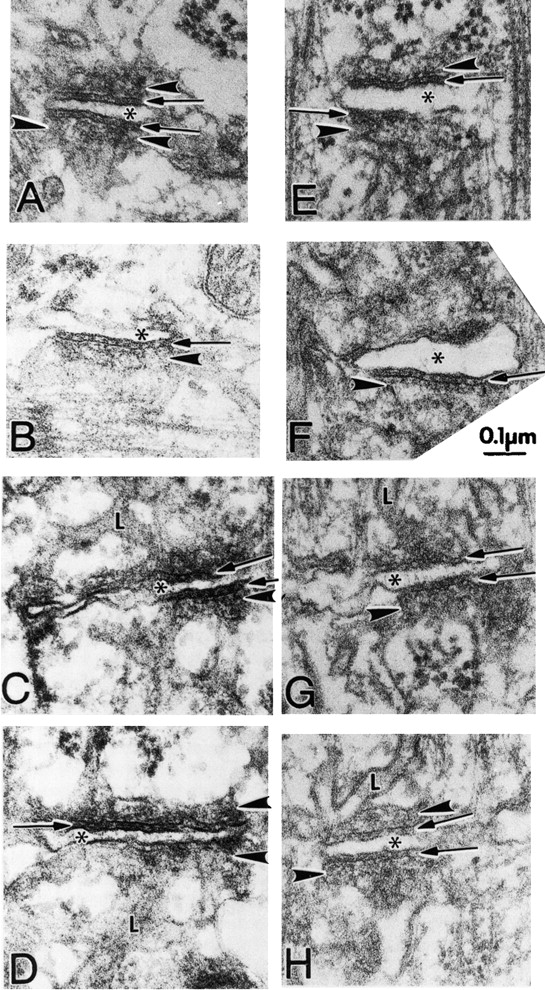
Triads appear structurally normal in ankyrin-B (−/−) skeletal muscle. Electron micrographs (see Materials and Methods) comparing T-tubule–SR junctions (triads) of mouse skeletal muscle from the quadriceps of normal (A–D) and ankyrin-B (−/−) (E–H) littermates at the same magnification. In both wild-type and ankyrin-B (−/−) fibers, the junctional SR (JSR) is seen more or less en face. The JSR membrane is usually marked by at least two rows of particles parallel to the junction and is frequently marked by filamentous densities extending away from the junction at right angles. The rows of particles can be seen between the arrowheads in A and opposite the arrowheads in B, C, E, F, and H. The filamentous density is best seen between arrowheads in A and opposite lower arrowheads in B, E, and F. In H, the two rows of particles appear as a jagged line (arrowhead), whereas in D and G, density on the JSR appears amorphous. Normal junctions show periodic foot processes in the gap (arrows) between T-tubule (*) and junctional SR membranes (arrowheads). Ankyrin-B (−/−) T–SR junctions show some densities between the SR and T-tubule membranes, particularly clear in (F), but the images in (E) and (H) are more common. The JSR is continuous with the tubular network of longitudinal SR (L) in both mutant and wild-type (see C, G, D, and H).