
Figure 8.
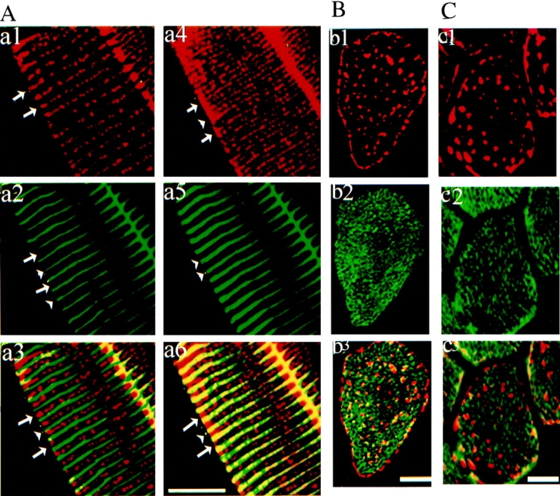
Ankyrin-B in skeletal muscle is localized at costameres and intracellular sites that are distinct from localization of SERCA 1 and ryanodine receptor type 1. (A). Immunolabeling of a longitudinal section of skeletal muscle with rabbit antibody against ankyrin (a1, a4, red) and a mouse monoclonal antibody against α-actinin (a2, a5, green) with a composite image (a3 and b3). Optical sections were taken at two different Z-heights: at the top, tangential to the cell surface (Z = 0 μm; a4–a6) and in the mid-region of the same cell (Z = −5 μm; a1–a3). Note that arrowheads point to location of Z line (α-actinin) and long arrows to the A-band. Note that ankyrin B labeling aligned with the Z-line appears just at the plasma membrane level (right). B and C show immunofluorescence labeling of cross-sections of skeletal muscle with rabbit antibody against the ankyrin-B (red, b1, b3, c1, and c3) and antibody against either SERCA 1 (B, green, b2 and b3) or ryanodine receptor type 1 and 2 (C, green, c2 and c3). Bars, 10 μm.