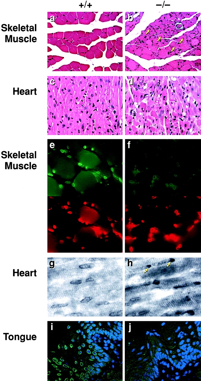
Figure 4.

Histological and immunohistochemical analysis of tissues from Lmna null mice. (a) Wild-type perivertebral muscle showing the peripheral localization of the nuclei in the muscle fibers. (b) Perivertebral muscles from Lmna null mice showing an increase in nuclear number with many centrally located within the muscle fibers (arrows). (c) Wild-type ventricular cardiac muscle. (d) Ventricular muscle from a Lmna null mouse. (e and f) Emerin (upper panels) localization in the skeletal nuclei of wild-type (e) and lamin −/− (f) mice. In the −/− muscle, the signal intensity of emerin in the NE is weaker than in the +/+ nuclei. Anti–lamin B labeling is unchanged (lower panels rhodamine label). (g and h) Emerin staining in the cardiac nuclei of wild-type (g) and lmna −/− (h) mice. Note the polar distribution (arrows) of emerin in the −/− cardiac muscle NEs. (i and j) Emerin staining in the tongue epithelium. In wild-type epithelium, emerin localization to the NE is readily detectable (i), whereas in the lamin null mice it is lost (j). Nuclei i and j are costained with DAPI.
