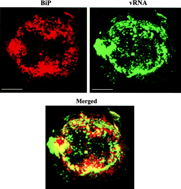
Figure 7.
vRNA colocalizes with BiP (ER marker). Protoplasts infected with wt vRNA were fixed at early stages of infection and processed for immunofluorescence using anti–BiP antibody and in situ hybridization. Most of the BiP (red) was associated with sites that contain vRNA (green). Merging the two images demonstrates colocalization of the signals (yellow in merged image). Bars, 10 μm.

