Figure 1.

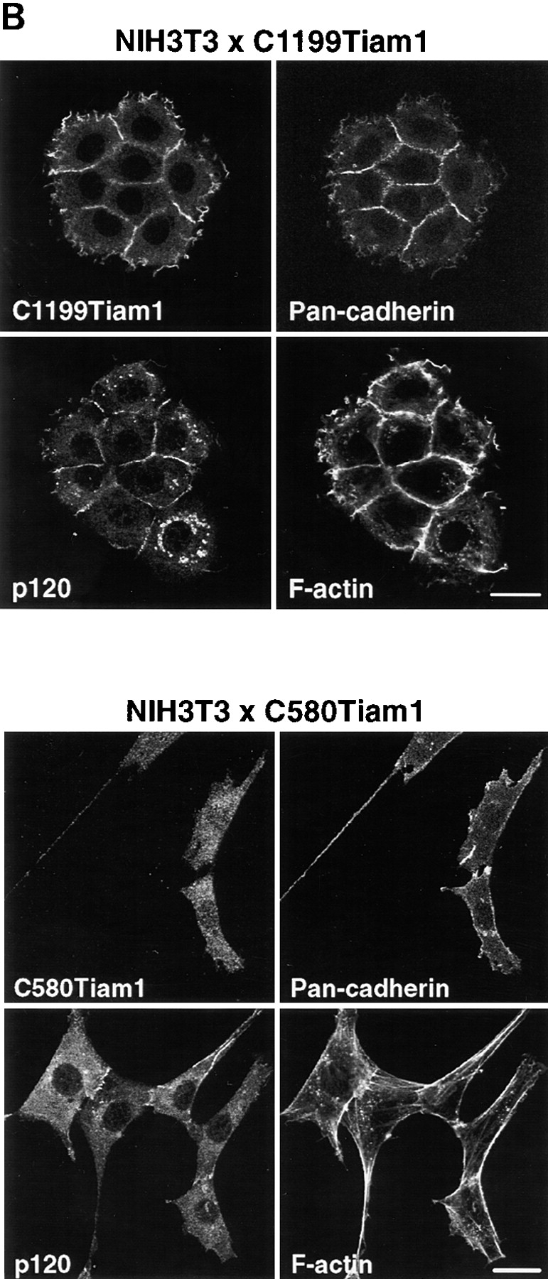
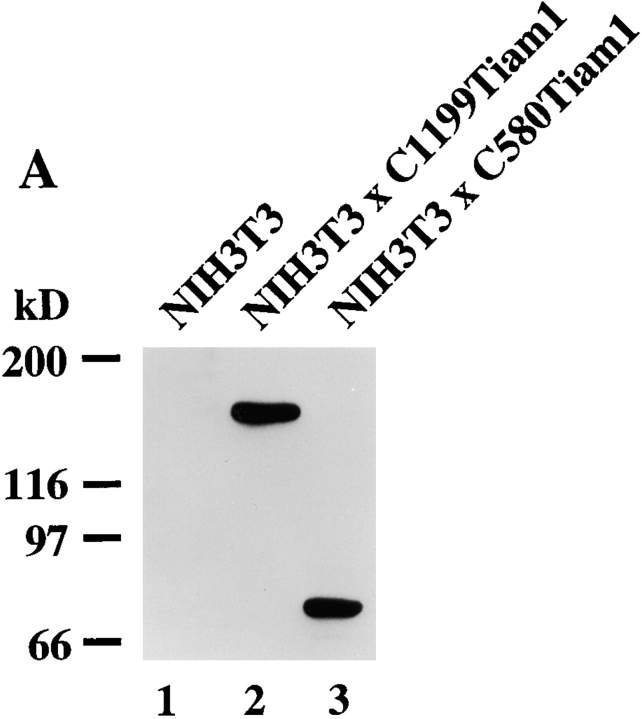
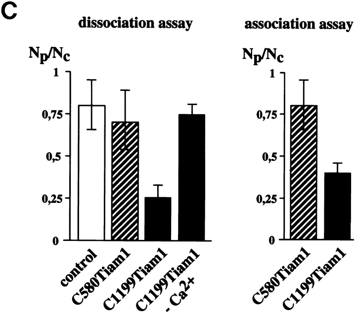
C1199Tiam1 induces an epithelial-like morphology in NIH3T3 cells by increasing N- and P-cadherin-based cell–cell adhesion. (A) Western blot using the Tiam1-specific C16 antibody of C1199- or C580Tiam1 immunoprecipitated with anti–HA antibody from the respective stable NIH3T3 cell lines. (B) NIH3T3 cells stably expressing C1199- or C580Tiam1 were stained for Tiam1, N- and P-cadherin (stained with a Pan-cadherin antibody), p120CAS, and F-actin. C1199Tiam1 localizes to sites of cell–cell contact and to membrane ruffles, whereas C580Tiam1 localizes to the cytoplasm. Cadherins as well as p120CAS localize to sites of cell–cell contact that were induced by C1199Tiam1. Bars, 25 μm. (C) Quantification of cell–cell adhesion of control NIH3T3 cells and cell lines expressing C1199- or C580Tiam1 determined by dissociation (in the presence and absence of Ca2+) and association assays and expressed as numbers of particles (cell clusters) per total number of cells (Np/Nc). Each bar represents the mean ± SD of triplicate assays. Shown is one representative example of three independent experiments.


