
Figure 1.
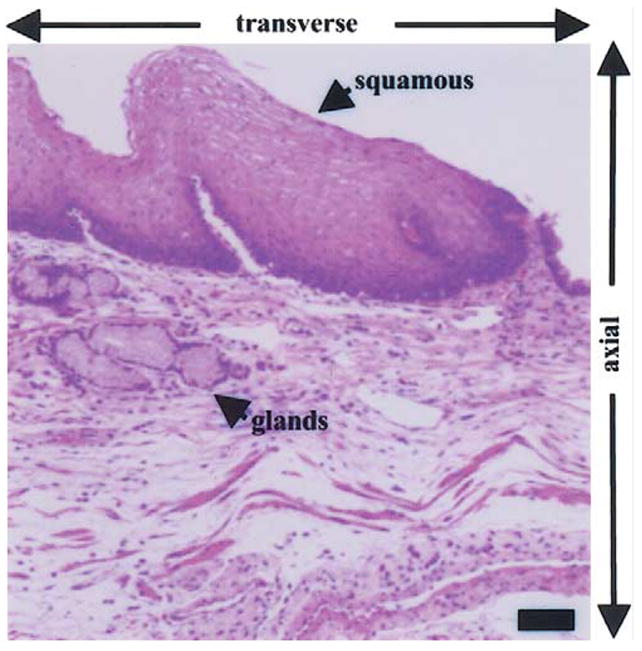
Histological examination of esophagus shows the presence of glands below the squamous epithelium, illustrating the need for optical methods to evaluate below the mucosal surface and for sub-cellular resolution in the axial and transverse dimensions. Scale bar, 50 μm. (Hematoxylin and eosin.)