
Figure 11.
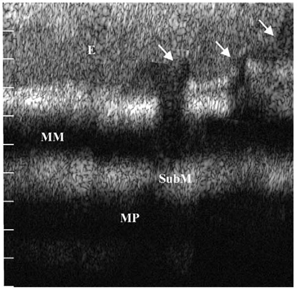
Dual-axes confocal image of the neosquamocolumnar metaplastic junction of resected human esophagus shown over a depth of 1 mm shows intact squamous epithelium (E), muscularis mucosa (MM), submucosa (SubM), and muscularis propria (MP) on the left and columnar mucosa with pit epithelium (arrows) on the right. Reprinted with permission.58