
Figure 12.
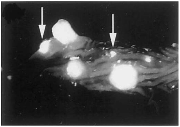
Near-infrared fluorescence image of an excised specimen of the colon from an APC min mouse after injection with the cathepsin B probe. Several 2–5-mm diameter polyps and adenomas as small as 50 μm (arrows) can be seen. Reprinted with permission.71