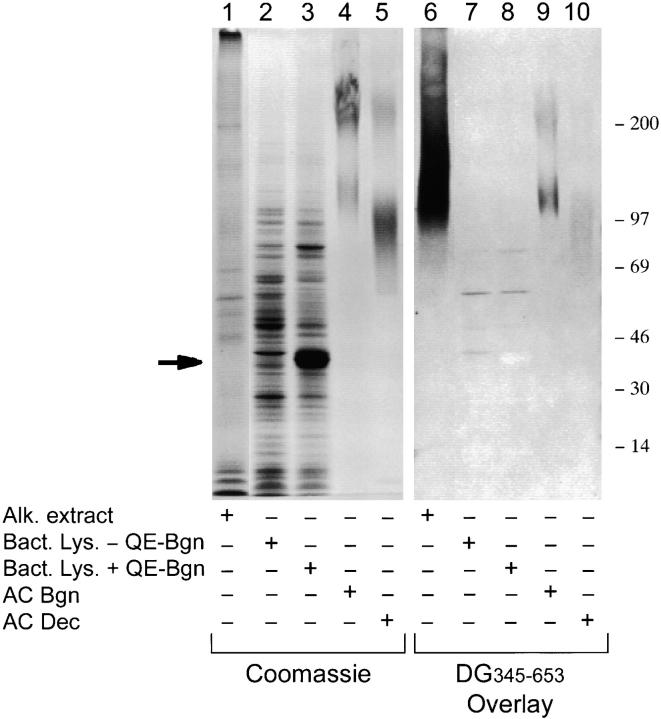
Figure 5.
The binding of dystroglycan to biglycan is dependent upon specific chondroitin sulfate side chains. Biglycan (or decorin) was analyzed by SDS-PAGE and Coomassie Brilliant blue staining for protein (lanes 1–5), or blot overlay assay for dystroglycan binding (lanes 6–10). Lanes 1 and 6, alkaline extract of Torpedo synaptic membranes (1 μg total protein); lanes 2 and 7, lysate of noninduced bacteria; lanes 3 and 8, lysate of induced bacteria expressing recombinant human biglycan (QE-Bgn; prominent band at ∼37 kD, arrow); lanes 4 and 9, biglycan purified from bovine articular cartilage (4 μg; Sigma Chemical Co.); lanes 5 and 10, decorin purified from bovine articular cartilage (4 μg; Sigma Chemical Co.). Biglycan present in electric organ binds dystroglycan much more strongly then biglycan or decorin purified from articular cartilage (compare Coomassie staining to dystroglycan overlay). Note that 4 μg of purified biglycan are present in lanes 4 and 9, compared with only 1 μg of total protein in lanes 1 and 6, of which biglycan is estimated to be <2%.

