Figure 7.
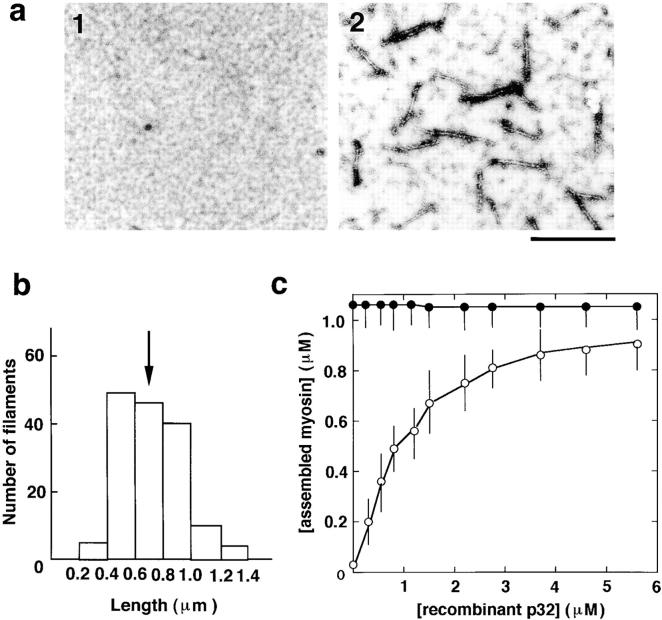
Myosin assembly by human p32. a, Electron micrographs of 0.2 μM unphosphorylated myosin alone (1) or mixed with 0.6 μM human p32 (2). b, Length distribution of myosin filaments formed by human p32. The histogram was obtained by measurement of 150 filaments. An arrow indicates average length. c, Quantification of myosin assembly by the sedimentation assay. Myosin at 1.1 μM was mixed with human p32 and subjected to the centrifugation assay of assembly. Amounts of assembled myosin were plotted against concentration of human p32. Open and closed circles denote unphosphorylated and phosphorylated myosins, respectively.

