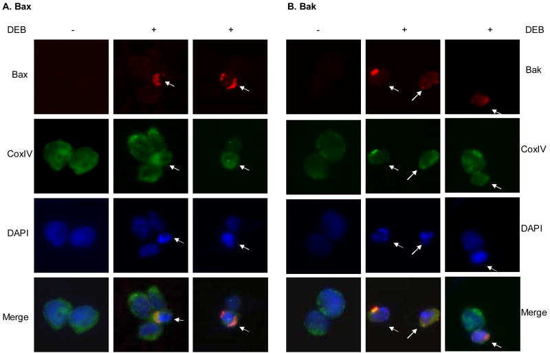
Figure 2. Localization of activated Bax and Bak proteins in TK6 cells after DEB treatment.
Cells were exposed to vehicle or 10 μM DEB for 14 h, fixed, permeabilized, and subjected to immunofluorescence analysis using antibodies specific for Bax (panel A, red) and Bak (panel B, red) proteins in active conformation. Mitochondrial identification was performed by staining with an Alexa-Fluor 488-conjugated mouse antibody against the mitochondria marker CoxIV (green). Nuclei were counterstained with DAPI (blue). Fluorescence from the various samples was detected by using a Nikon E400 fluorescence microscope. The same microscopic field for each sample was analyzed for all three fluorochromes, and an overlay of all three microscopic fields is also shown (as merge). Arrows point to cells undergoing early stage DEB-induced apoptosis, as identified by their condensed nuclei after DAPI staining.

