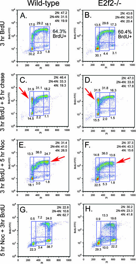
FIG. 7.
E2f-2 loss is associated with both a delay in S-phase progression and a failure to maintain mitotic arrest. (A to H) Representative BrdU labeling and propidium iodide (PI) staining of E13.5 wild-type (A, C, E, and G) or E2f2−/− (B, D, F, and H) fetal liver erythroblasts cultured for 3 h in the presence of BrdU to assess the cell cycle ex vivo (A and B); cultured for 3 h in the presence of BrdU, followed by 5 h in the absence of BrdU, to assess cell cycle progression to BrdU-positive G1 (C and D); cultured for 3 h in the presence of BrdU, followed by 5 h in the presence of nocodazole (Noc), to assess cell cycle progression to BrdU-positive G2/M and maintenance of a mitotic arrest (E and F); and cultured in nocodazole for 5 h to arrest cells at mitosis, followed by the addition of BrdU for 3 h of incubation, to assess S-phase entry, S-phase clearance to G2/M, and maintenance of a mitotic arrest (G and H).

