FIG. 2.
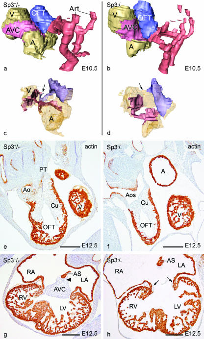
Morphological analysis of Sp3−/− embryonic hearts. (a to d) 3D reconstructions of a WT (a and c) and an Sp3−/− (b and d) embryonic heart at E10.5. Color codes: light brown, atria (A) and ventricles (V); pink, AVC; blue, outflow tract cushions (OFT); red, arteries (Art). The outer surface of the heart (a and b) shows the different relative positions of the cardiac segments in the Sp3−/− embryo. In the WT heart, the AVC and OFT cushions meet at the inner curvature (arrow in panel c), while in the knockout there is a wide inner curvature and separation of the cushions (arrow in panel d). (e to h) Sections of a WT (e and g) and an Sp3−/− (f and h) heart, stained for actin, show at E12.5 that in the Sp3−/− embryo the septation of the aortic sac (Aos) is not yet complete (f) compared to in the normal embryo (e), where the aorta (Ao) and pulmonary trunk (Pt) are already separated. At the atrioventricular canal level (g) in the WT, the AVC tissue is already fused with the cushion tissue (arrowhead) at the base of the atrial septum (AS). In the mutant heart (h), a common valve is shown. Small lateral cushions are present (arrows), and the cushion tissue at the base of the AS is not connected to the remaining AVC tissue (small patch in the middle). Bars, 200 μm.

