
Figure 2.
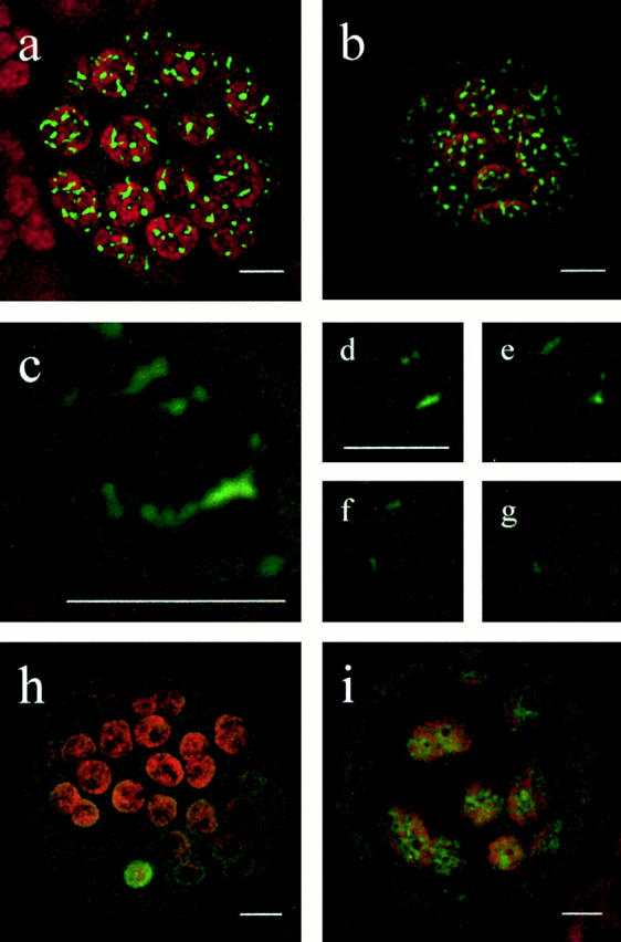
Subcellular localization of FtsZ-GFP fusion proteins. Physcomitrella protoplasts were transiently transfected with the corresponding expression plasmid and GFP fluorescence was visualized by CLSM 2 d after transfection. (a) Protoplast transfected with PpFtsZ1-GFP. (b) Protoplast transfected with PpFtsZ2-GFP. (c) Single chloroplast (detail of a). (d–g) Sections of the chloroplast shown in c demonstrating the localization of PpFtsZ1 solely within chloroplasts. (h) Protoplast transfected with pFtsZ1(1–35)-GFP (incomplete transit peptide) showing cytoplasmic distribution of fusion protein. (i) Protoplast transfected with pFtsZ1(1–93)-GFP (complete transit peptide) showing plastidic distribution of fusion protein. a–c, and h and i, represent overlays of all sections of the object. Chlorophyll fluorescence is shown in red; GFP signals are shown in green. Bars, 5 μm.