
Figure 9.
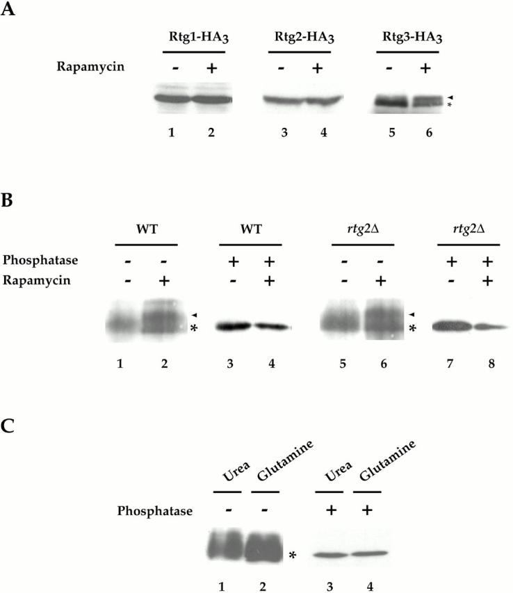
Rtg3 is a phosphoprotein and is differentially phosphorylated after rapamycin treatment. (A) Cells expressing Rtg1-HA3 (PLY047), Rtg2-HA3 (PLY089), and Rtg3-HA3 (PLY050) were grown to 0.5 OD600/ml in YPD and were treated either with drug vehicle alone or with rapamycin for 15 min. Extracts were prepared and Western blot analysis was performed using anti–HA monoclonal antibodies to detect each protein. No change in the abundance or relative mobility of Rtg1 or Rtg2 could be detected after rapamycin treatment. In contrast, a portion of Rtg3 showed an increased mobility (arrowhead) after rapamycin treatment, compared with its mobility in the absence of rapamycin (*). (B) Wild-type (K699) and rtg2Δ (EY0734) cells transformed with pRtg3-zz and were grown to 0.5 OD600/ml in SCD media lacking uracil. Cells were then treated with drug vehicle or with rapamycin for 15 min. Extracts were prepared and Rtg3-zz was immunoprecipitated with IgG-Sepharose and either mock-treated or treated with phosphatase before Western blot analysis, as indicated. Increased mobility of a portion of Rtg3-zz after rapamcyin treatment is indicated (arrowhead). (C) Wild-type (K699) cells carrying pRtg3-zz were grown to 0.5 OD600/ml in MD-glutamine or MD-urea and processed as in B. For each experiment in A–C, all samples were from the same gel. Identical results were obtained in three separate experiments.