
Figure 2.
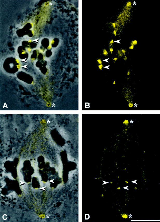
Dynein staining in grasshopper spermatocytes. (A and C) Superimposed phase–contrast and immunofluorescence images. (B and D) Immunofluorescence images. The dynein antibody stains the spindle, spindle poles, and kinetochores (arrowheads) in both prometaphase (A and B) and metaphase (C and D) cells (the absence of a tight metaphase plate in C and D is due to cell lysis before fixation). Dynein staining is far brighter on prometaphase kinetochores (A and B) than at metaphase (C and D). *Spindle poles. Bar, 10 μm.