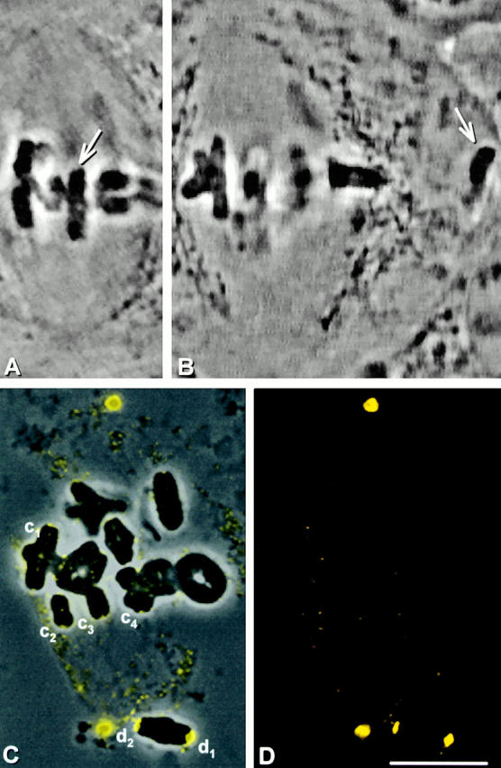
Figure 5.
Dynein staining at kinetochores after microtubule disassembly. (A and B) Live cell images. A chromosome (arrow) was detached from a metaphase spindle, placed in the cytoplasm, and held there for 10 min. The cell was then lysed in the presence of a calcium-containing microtubule disassembly medium. (C) Superimposed phase–contrast and immunofluorescence images. (D) Immunofluorescence image. (E) Composite showing the kinetochores labeled in C at higher magnification. The kinetochores that had been detached for 10 min (d1 and d2) are brighter than the kinetochores that remained attached until calcium treatment (c1–c4). Bars, 10 μm.