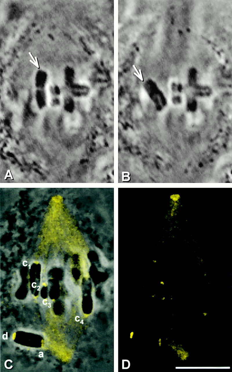
Figure 6.
The consequence of releasing tension, but not microtubule attachment, at kinetochores. (A and B) Live cell images. One kinetochore (arrow) of a chromosome was detached from a metaphase spindle and kept detached for 10 min. (C) Superimposed phase–contrast and immunofluorescence images. (D) Immunofluorescence image. (E) Composite showing the kinetochores labeled in C at higher magnification. The detached kinetochore (d) stained brightly for dynein, whereas the kinetochore that was relaxed, but still attached (a) is only as bright as kinetochores that remained under tension (c1–c4). Bars, 10 μm.