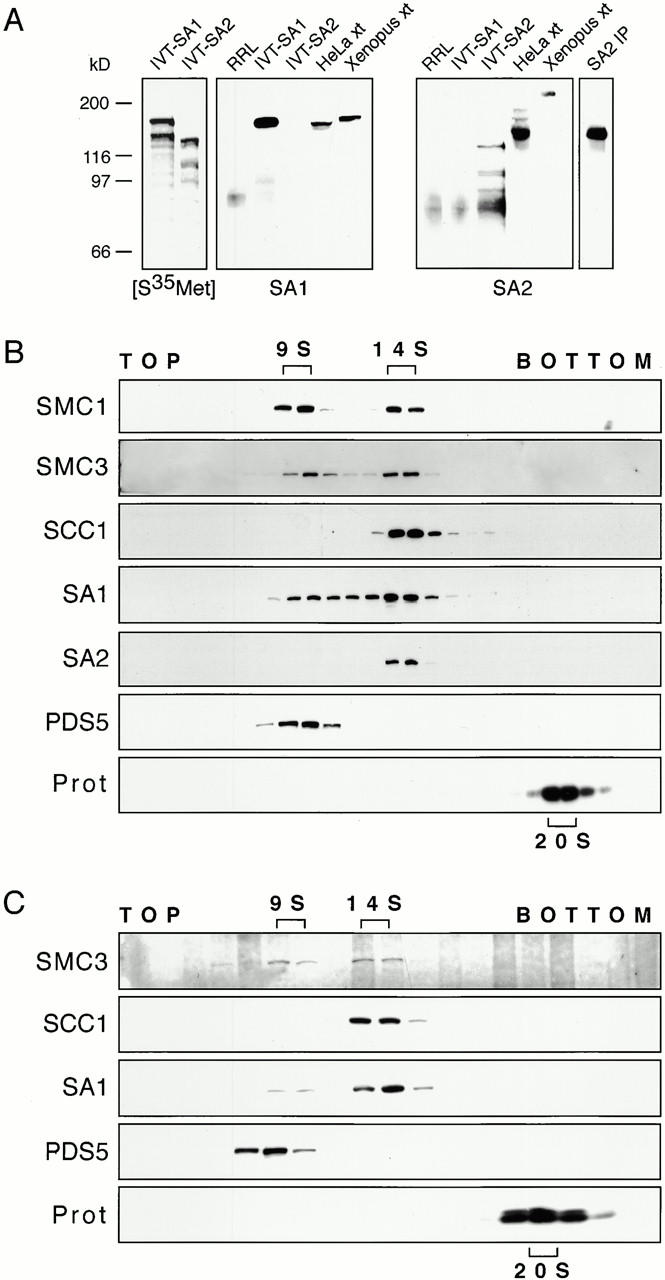
Figure 1.

Fractionation of human cohesin complexes by sucrose density gradient centrifugation. (A) Characterization of SA1 and SA2 antibodies. (Left) PhosphorImager scan of in vitro–translated 35S-labeled human SA1 and SA2 (IVT-SA1, IVT-SA2) separated by SDS-PAGE. Other panels, control rabbit reticulocyte lysate (RRL), in vitro–translated SA1 and SA2, protein extracts (xt) from HeLa cells, and Xenopus interphase egg extracts, and SA2 (446) immunoprecipitates isolated from HeLa extracts (SA2 IP) were analyzed by SDS-PAGE and immunoblotting with specific SA1 (444) or SA2 (446) antibodies. (B) Sucrose gradient fractions containing proteins from logarithmically growing HeLa cells were analyzed by SDS-PAGE and immunoblotting with antibodies to the indicated proteins. SA1 and SA2 were detected with antibodies 444 and 446, respectively. Prot, proteasome. (C) Sucrose gradient fractions containing proteins from Xenopus interphase extract were analyzed by SDS-PAGE and immunoblotting with antibodies to the indicated proteins.
