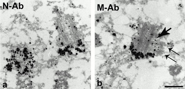
Figure 6.
Immunoelectron microscopic localization of C-Nap1 domains. U2OS cells were fixed with 3% paraformaldehyde/2% sucrose and subjected to immunoelectron microscopic analysis with silver-enhanced Nanogold®. Both N-Ab (a) and M-Ab (b) strongly labeled the proximal ends of both centrioles. Note that no C-Nap1 staining could be seen at the interface (b, arrow) between procentriole (b, thin double arrows) and centriole. Bar, 250 nm.

