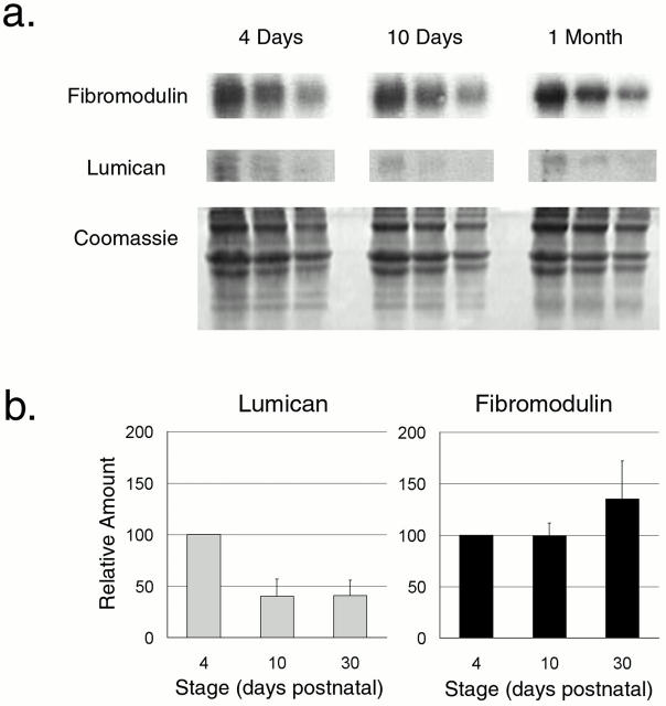
Figure 3.
Lumican and fibromodulin content during normal tendon development. (a) A representative semiquantitative Western analysis of lumican and fibromodulin during development in the normal mouse tendon is presented. Tendons were extracted in 4 M guanidine-HCl at 4 d, 10 d, and 1 mo. 80, 40, and 20 μg of total protein from each time point were loaded onto the gel. The core proteins were transferred, reacted with antilumican or antifibromodulin antisera followed by radiolabeled goat anti–rabbit IgG, and quantitated using phosphoimaging. A duplicate gel stained with Coomassie shows similar amounts of type I collagen in the extracts. (b) The relative lumican and fibromodulin content in the tendon at 4 d, 10 d, and 1 mo postnatal were derived from three independent experiments. The mean values for both lumican and fibromodulin were set to 100 at 4 d and the results were plotted as a function of development (bars indicate SD).

