
Figure 5.
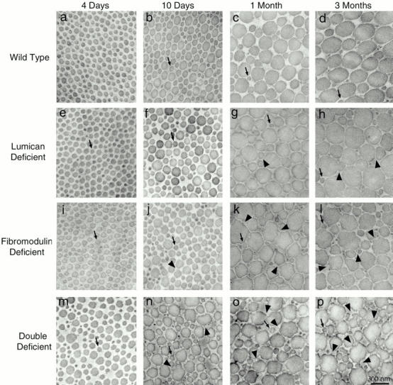
Collagen fibril structure during development in normal wild-type, lumican-, fibromodulin-, and double lumican/fibromodulin–deficient mice. Transmission electron micrographs of transverse sections from mouse flexor tendons from normal mice (a–d) and mutant mice (e–p). Fibril structure was analyzed at different developmental stages between 4 d and 3 mo postnatal, 4 d (a, e, i, and m), 10 d (b, f, j, and n), 1 mo (c, g, k, and o), and 3 mo (d, h, l, and p) postnatal. Arrows indicate fibrils with diameters of ∼64 nm, the diameter seen in normal 4-d postnatal tendons. Arrowheads indicate irregular fibril profiles. Bar, 300 nm.