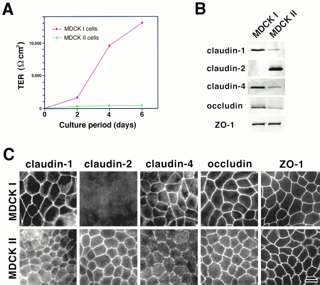
Figure 1.
Claudins in MDCK I and II cells. (A) TER measurement of MDCK I and II clones used in this study. MDCK I or II cells (4 × 10⁵ cells) were plated on 24-mm filters. In 6-d culture, the TER values of MDCK I and II cells reached the maximum levels, 12,992 ± 594 and 206 ± 35 Ωcm2, respectively (mean ± SD, n = 11). (B) Immunoblotting. Total lysates of MDCK I and II cells were separated by SDS-PAGE, followed by immunoblotting with pAbs for claudin-1, -2, and -4, and mAbs for occludin and ZO-1. In both MDCK I and II cells, claudin-1 and -4 were expressed, although their expression levels in MDCK II cells were significantly lower than those in MDCK I cells. Claudin-2 was expressed only in MDCK II cells. Occludin was expressed in larger amounts in MDCK I than MDCK II cells. (C) Immunofluorescence microscopy. In MDCK I cells, claudin-1, claudin-4, occludin, and ZO-1 were coconcentrated at TJs, where claudin-2 was undetectable. In contrast, in MDCK II cells, in addition to claudin-1, claudin-4, occludin, and ZO-1, claudin-2 was clearly detected at TJs. The claudin-4 signal was weak. Bar, 10 μm.

