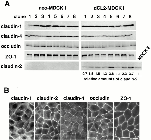
Figure 3.
Establishment of MDCK I transfectant clones stably expressing claudin-2. Dog claudin-2 and neomycin-resistance genes were introduced into MDCK I cells, and eight independent clones for each were established (dCL2-MDCK I and neo-MDCK I, respectively). (A) Immunoblotting. Total lysates of neo-MDCK I and dCL2-MDCK I clones (and also MDCK II cells) were subjected to SDS-PAGE in the same amount of total proteins, followed by immunoblotting. There were no significant differences in the expression levels of claudin-1, claudin-4, occludin, or ZO-1, except for the dCL2-MDCK I–specific expression of claudin-2 between neo-MDCK I and dCL2-MDCK I clones. The amounts of endogenous claudin-2 in MDCK II cells as well as exogenous claudin-2 in dCL2-MDCK I cells were quantified as described in Materials and Methods, and their dCL2-MDCK I/MDCK II ratios (relative amounts of claudin-2) were calculated. (B) Immunofluorescence microscopy. In clone 2 of dCL2-MDCK I cells, together with endogenous claudin-1, claudin-4, occludin, and ZO-1, exogenously expressed claudin-2 was co-concentrated at TJs. Bar, 10 μm.

