
Figure 5.
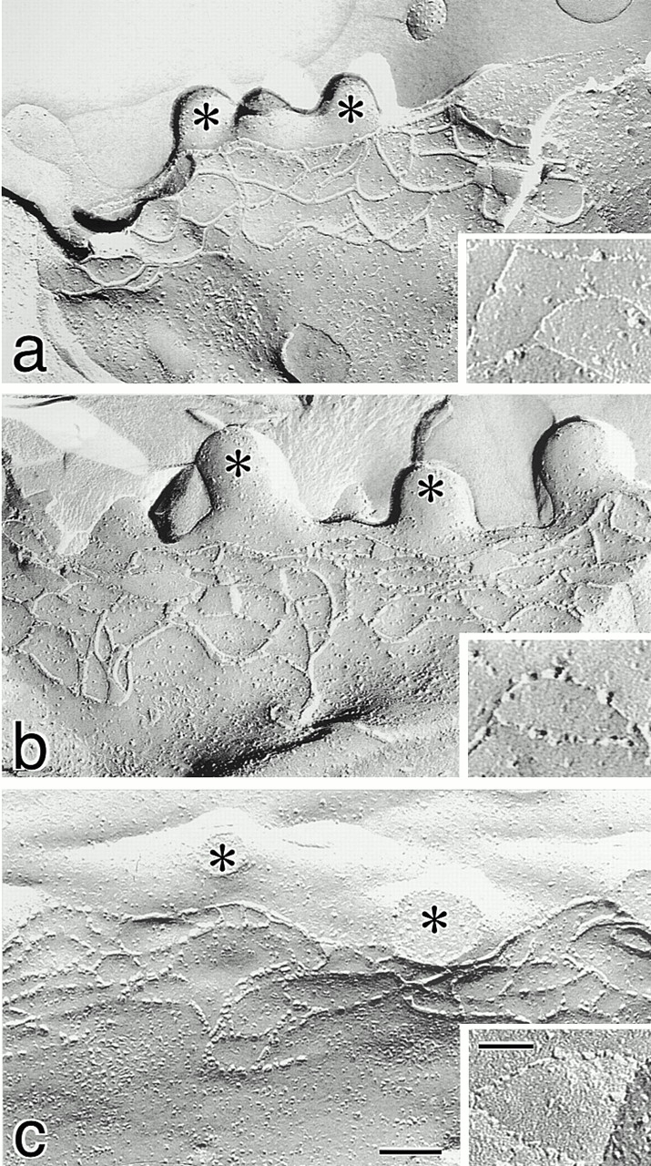
Freeze-fracture replica images of TJs in neo-MDCK I (a), dCL2-MDCK I (b), and MDCK II cells (c). Cells (4 × 10⁵ cells) were plated on 24-mm filters, cultured for 6 d, fixed with glutaraldehyde, and then processed for freeze-fracture replica electron microscopy. The number of TJ strands in neo-MDCK I cells was similar to those in dCL2-MDCK I as well as MDCK II cells (Table ), and the network pattern of TJ strands in neo-MDCK I cells did not appear to be more complex than that in dCL2-MDCK I or MDCK II cells. In neo-MDCK I cells (a), TJ strands were largely associated with the P-face and were mostly continuous, and on the E-face (inset) complementary continuous grooves were vacant. In dCL2-MDCK I cells (b), TJ strands were fairly discontinuous on the P-face, and on the E-face (inset) intramembranous particles were scattered within the grooves. The strands (c) and grooves (inset) of MDCK II cells were similar in appearance to those in dCL2-MDCK I cells. *Microvilli. Bar, 100 nm; inset, 50 nm.