
Figure 5.
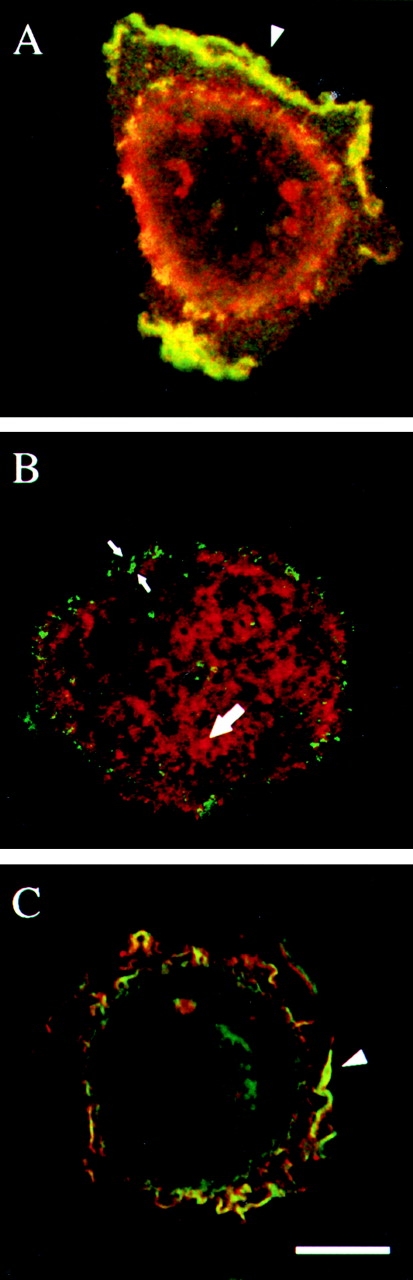
The α6β4 integrin colocalizes with the EGF receptor and phosphotyrosine in ruffles and lamellipodia but not in hemidesmosomes. A431 cells were plated on laminin-1 for 1 h and then either stimulated with EGF (1 ng/ml) for 15 min (A and C) or left untreated (B). All cells were processed for double-staining with the α6-specific rat mAb GoH3 (red) and mouse Abs specific for either the EGFR (A, green) or phosphotyrosine (PY20; B and C, green). Yellow depicts colocalization of two antigens. Arrowheads indicate colocalization in lamellipodia and ruffles, and small arrows indicate focal adhesion-like structures. Large arrow indicates hemidesmosome area. Bar, 10 μm.