Figure 5.
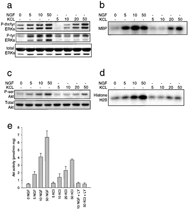
Activation of ERK and Akt kinases by NGF and KCl. a and b, ERK activation in response to increasing concentrations of NGF or KCl. a, Western blot analysis of equal amounts of protein derived from sympathetic neurons treated for 15 min with increasing concentrations of NGF (ng/ml) or KCl (mM), as detected with antibodies to tyrosine/threonine-phosphorylated ERKs (P-tyr/thr ERKs), tyrosine-phosphorylated ERKs (P-tyr ERKs), or to total ERK protein (total ERKs). The upper band in the doublet is ERK1 and the lower is ERK2. Note that all three panels are reprobes of the same blot. b, In vitro ERK activation assay using myelin basic protein (MBP) as a substrate in lysates of sympathetic neurons treated for 15 min with increasing concentrations of NGF (ng/ml) or KCl (mM). All samples were normalized for equal amounts of protein. c–e, Akt activation in response to increasing concentrations of NGF or KCl. c, Western blot analysis of equal amounts of protein derived from sympathetic neurons treated for 15 min with increasing concentrations of NGF (ng/ml) or KCl (mM), as detected with antibodies to serine-phosphorylated Akt (P-ser Akt) or to total Akt protein (Total Akt). Note that both panels are reprobes of the same blot. d, In vitro Akt activation using histone H2B as a substrate in lysates of sympathetic neurons treated for 15 min with increasing concentrations of NGF (ng/ml) or KCl (mM). All samples were normalized for equal amounts of protein. e, Quantitative in vitro Akt activity assay using an Akt-specific substrate in lysates of sympathetic neurons treated for 15 min with increasing concentrations of NGF (ng/ml) or KCl (mM). In sister cultures, neurons were coincidently treated with 100 μM of the PI3-kinase inhibitor LY294002 (LY). Bars represent the mean ± SD. Note that inhibition of PI3-kinase with LY294002 abolishes Akt activity induced by NGF or KCl.

