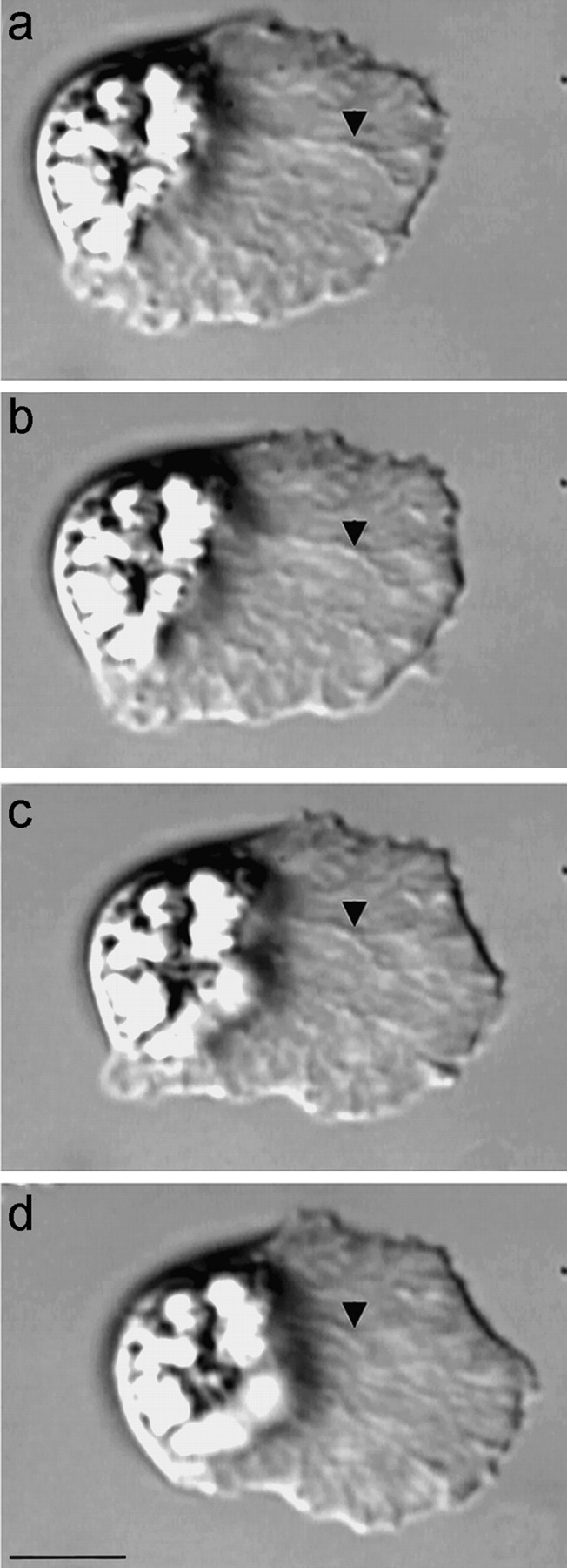
Figure 1.

MSP cytoskeletal dynamics in crawling Ascaris sperm. The long branched elements that extend from the leading edge to the base of the lamellipodium are the fiber complexes, each a dense meshwork of MSP filaments. The cytoskeleton flows retrograde as the fiber complexes are assembled at the leading edge and disassembled at the cell body. Because the rates of cytoskeletal flow and locomotion are coupled, morphological markers in the cytoskeleton, such as the branch in the fiber complex indicated by the arrowhead, remain nearly stationary relative to the substrate. The fields of view are identical in each frame and the interval between frames is 10 s. Over the 30-s interval from a–d, both the leading edge and the cell body advanced by 6.5 μm while the lamellipodium maintained a length of 25 μm. This illustrates the balance between the rates of cytoskeletal assembly/leading edge protrusion and cytoskeletal disassembly/cell body retraction during sperm locomotion. Bar, 10 μm.
