
Figure 3.
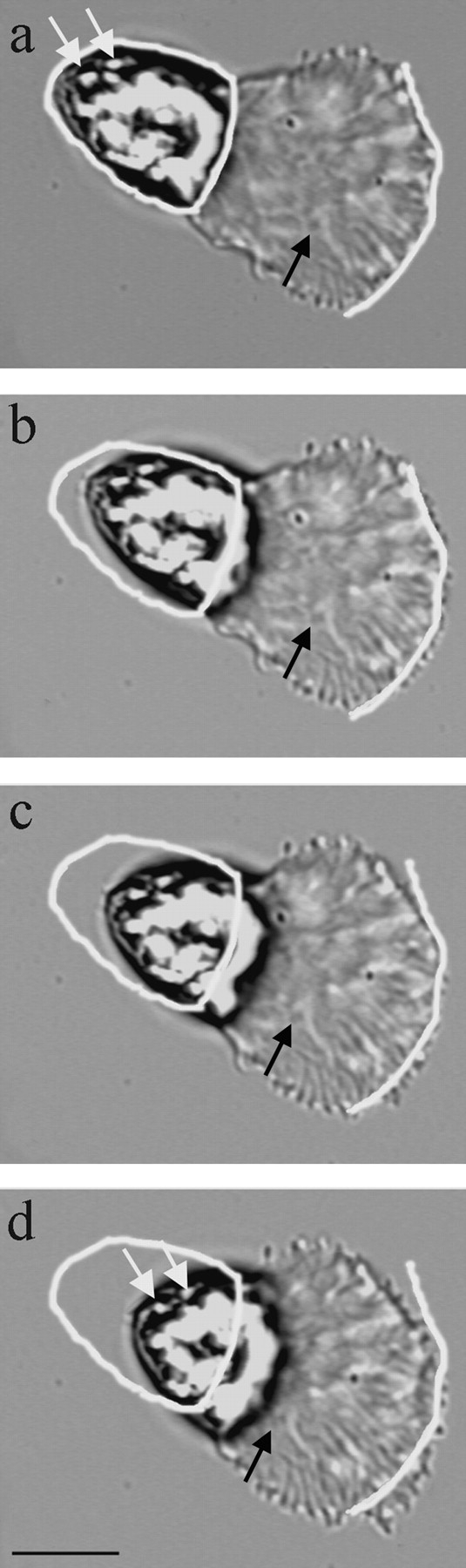
Treatment of sperm with acetate buffer, pH 6.75, uncouples protrusion from retraction. The positions of the cell body and the leading edge, when the cell was perfused with acetate buffer (a), are outlined in white in a–d. During this sequence (elapsed time, 30 s) part of the leading edge protruded slowly (3 μm/min; b–d) while an adjacent portion, toward the top of the frame (c), retracted slightly. Thus, treatment with acetate buffer slowed cytoskeletal assembly dramatically. However, cytoskeletal disassembly was not inhibited and so the lamellipodium and the fiber complexes within shortened and the cell body continued to move forward at 15 μm/min. The black arrow indicates a kink in a fiber complex that moved rearward with respect to the leading edge during the sequence. This indicates that cytoskeletal treadmilling persists even when the rate of protrusion slowed. Note that the distance between the cell body and this kink decreases throughout the sequence, due to continued disassembly at the base of the lamellipodium. The white arrows indicate refringent spots that maintained their position in the cell body during retraction. This shows that the cell body moves forward without rolling. Interval between frames, 10 s. Bar, 10 μm.