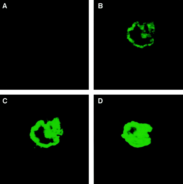
Figure 5.
Confocal microscopic localization of the CD38 antigen on the osteoclast plasma membrane. Intense peripheral immunofluorescent staining of osteoclasts with a highly specific agonist anti-CD38 monoclonal antibody, A10. 1-μm-thick serial sections in the coronal plane (B–D) are shown. Note that osteoclasts incubated with no antiserum (not shown), nonimmune mouse IgG (not shown), or an irrelevant antibody (Ab34) (A) do not stain. For details on confocal microscopy, see Materials and Methods.

