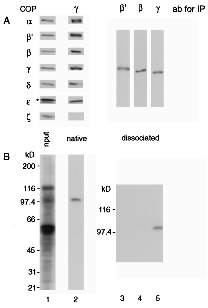
Figure 3.
Photocrosslinking of cell lysate with 125I-F*-p23wt in solution. CHO cell lysate was incubated with 125I-F*-p23wt in solution for 1 h on ice prior to photoactivation for 2 min on ice. (A) Lane 1, immunoblot of total cell lysate analyzed with a mixture of antibodies against all seven coatomer subunits. The asterisk indicates a cross-reactivity of the anti-ɛ-COP antiserum with a protein present in the lysate. Lane 2, immunoblot analysis of coatomer immunoprecipitated with an anti-γ-COP antibody without prior dissociation. Lanes 3–5, immunoblot analysis of β′-, β-, and γ-COPs individually immunoprecipitated (IP) upon dissociation of coatomer by SDS treatment. Samples were separated on 7.5–16.5% (lanes 1 and 2) or 7.5% polyacrylamide gels (lanes 3–5) in the presence of SDS. (B) Autoradiogram of A.

