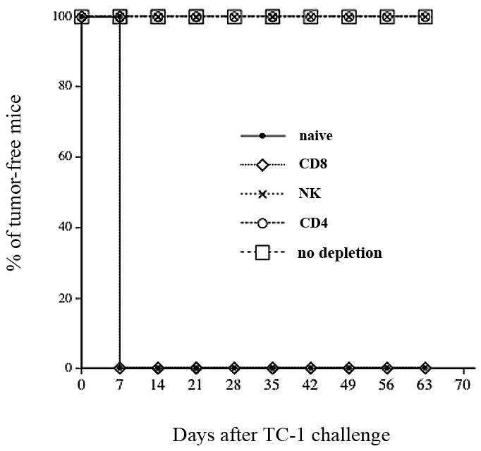
Figure 5. In vivo antibody depletion experiments.

Graphical representation of the percentage of tumor-free mice over time in each group of mice. C57BL/6 mice (5 per group) were immunized twice via gene gun with 2μg/mouse of pcDNA3-IL2-E7. One week after the last vaccination, the vaccinated mice were challenged subcutaneously with 5×10⁴ TC-1 cells/mouse. One week before the tumor challenge, the vaccinated mice were depleted of CD8, CD4 or NK cells using the 2.43, GK1.5 and PK136 monoclonal antibodies respectively every other day for 3 times for the first week and then once every week as described in the Materials and Methods section. The mice were monitored for evidence of tumor growth by inspection and palpation twice a week. The data shown here are from one representative experiment of two performed.
