
Figure 1. Compound C inhibits hypoxic activation of HIF-1.
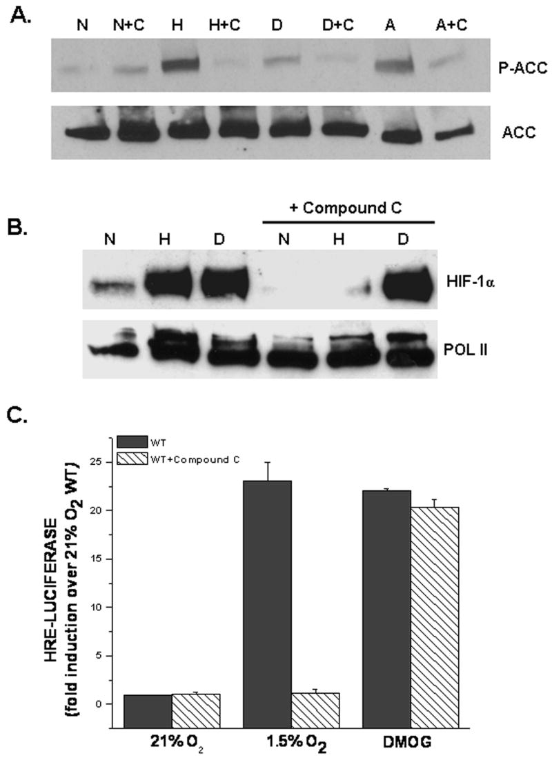
A. AMPK activation assessed by phospho-ACC protein in WT cells exposed to 21%O2 (N), 1.5%O2 (H), 1mM DMOG (D), or 0.5 mM AICAR (A) ± 20μM Compound C (C) for 30 mins. Anti-ACC antibody used as loading control.
B. HIF-1α protein levels in WT cells exposed to 21% O2 (N)± 1mM DMOG (D) or to 1.5% O2 (H) ± Compound C for 2 hours. Anti-POL II antibody used as loading control.
C. WT cells transfected with a HRE-Luciferase reporter gene construct and exposed to 21% O2 ± 1mM DMOG or to 1.5% O2 ± Compound C for 16 hours before being harvested. Firefly luciferase activity was normalized to renilla luciferase activity and data reported as fold induction over 21%O2.