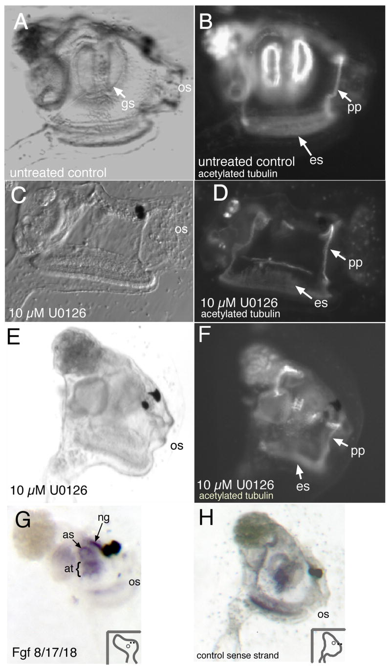
Fig. 3.
Inhibitor treatment disrupts siphon/gill slit formation in juveniles. (A, B) Control juveniles right-side view, oral siphon (os) is to the right. Post-metamorphosis animals show characteristic 1° gill slits. Staining for acetylated tubulin shows gills slits and peripharyngeal band (pp) and endostyle (es). (C, D) Early-treated animals which are allowed to recover and undergo metamorphosis do not develop gill slits. Tubulin staining is absent where gill slits normally form, but is still present in peripharyngeal band and endostyle. (E,F) Late treatment with U0126; Gill slits and atrial siphon are absent in the treated animal. Tubulin staining shows that while structures such as the peripharyngeal band and endostyle are present, well-formed gill slits are not. (G,H) Fgf8/17/18 in situ hybridization of juvenile Ciona savignyi. (G) Specific staining was observed around the atrial siphon opening (as), within the atrium (at), and in the neural gland (ng). (H) Control sense-strand probe does not show staining in neural gland or atrial siphon. (os, oral siphon.)

