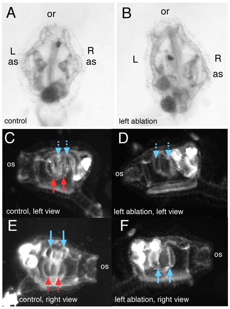
Fig. 5.

Laser ablation of an atrial siphon primordium disrupts later siphon and gill slit development. (A) Control, unablated juvenile, dorsal view, oral siphon (os) is at top. The chevron-shaped structure underlying right or left atrial siphons (as) are the primary gill slits. (B) A left-sided laser ablation of the atrial primordium at results in an animal with no atrial siphon and no primary gill slits on the ablated side. Left or right side views in dark field reveal paired primary gill slits on either side of a control animal (C,E); red arrows point to left-side primary gill slits, blue point to right-sided. Solid arrows are in the plane of focus; dashed arrows are slightly deeper, on the other side of the animal depicted in the panel. (D,F) A left-ablated juvenile reveals that primary gill slits form only on the untreated side.
