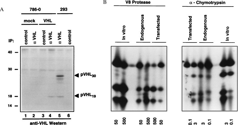
Figure 1.
Identification of pVHL19 in vivo. (A) 786-O VHL(−/−) renal carcinoma cells were transfected with plasmid encoding pVHL19 (lanes 3 and 4) or with the backbone expression plasmid (lines 1 and 2). The transfected cells and 293 human embryonic kidney cells (lines 5 and 6) were labeled with 35S, lysed, and immunoprecipitated with control or IG32 monoclonal anti-VHL antibody as indicated. Bound proteins were detected by immunoblot analysis with affinity purified polyclonal anti-VHL antibody. (B) The 35S-labeled pVHL19 bands corresponding to lanes 4 and 5 of A were excised and digested with the indicated amounts of α-chymotryspin (micrograms) and V8 protease (nanograms). pVHL19 translated in vitro was digested in parallel. Digestion products were resolved by SDS/PAGE and detected by autoradiography.

