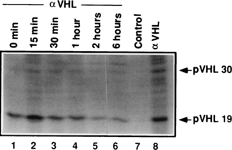
Figure 2.
Pulse-chase analysis of pVHL. 293 human embryonic kidney cells were pulse radiolabeled with [35S]methionine. Cell extracts were prepared at the indicated time points after chase with unlabeled methionine and immunoprecipitated with an anti-VHL mAb (IG32) under antibody excess conditions (lanes 1–6). In parallel, 293 cells were radiolabeled with [35S]methionine under steady-state conditions and immunoprecipitated with control (lane 7) or anti-VHL(IG32) (lane 8) antibody. Bound proteins were resolved by SDS/PAGE and detected by fluorography. Positions of pVHL30 and pVHL19, as confirmed by anti-VHL immunoblots performed in parallel, are shown by arrows.

