Figure 3.

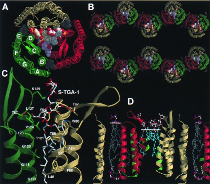
Lipid binding and layer packing of BR trimers in PM and monoclinic BR crystals. (A) Top view on the BR trimer/lipid complex from the extracellular side. Lipids are shown as space-filling models. Single phytanols (gray) are located on the cytosolic side of the BR trimer. (B) View along the a*-axis of the monoclinic BR crystal (monomer A, yellow; B, green; C, red). (C) Binding site of the glycolipid S-TGA-1 as viewed from the trimer axis. (D) Cross section of the PM model. Monomer B and an associated S-TGA-1 lipid replaced BR and the lipids 261, 266 of the previous PM model of Grigorieff et al. (11). The white lines show the head group/lipid boundaries.
