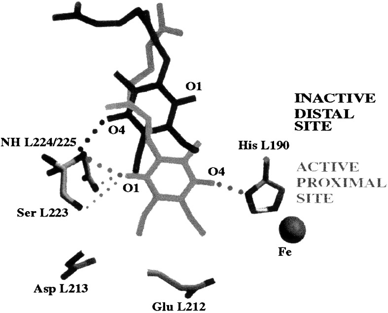
Figure 6.
Comparison of the binding positions for QB determined from the light (D+QA QB−⋅) and dark (DQAQB) x-ray crystal structures of the RC (10). Movement from an inactive–distal (black) to an active–proximal (gray) binding site is proposed as the major structural change involved with conformational gating of electron transfer from QA−⋅ to QB. Hydrogen bonding partners are connected by dotted lines. Modified from ref. 10.

