Figure 3.
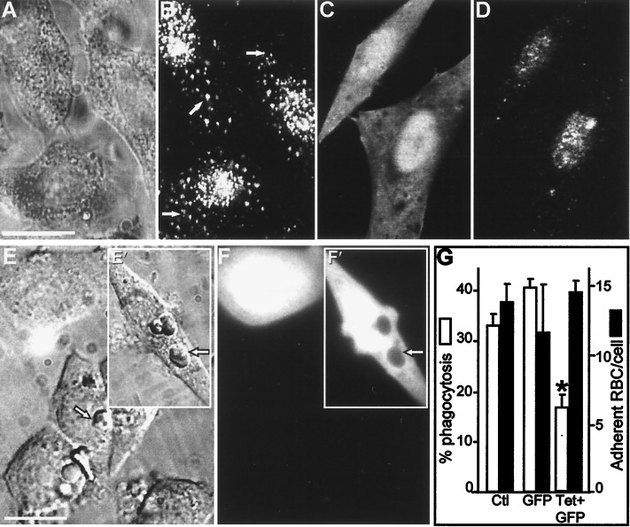
Impaired phagocytosis after degradation of VAMP-2 in FcγRIIA-transfected CHO cells. (A and B) VAMP-2 immunostaining of CHO cells. (A) Nomarski image. (B) VAMP-2 immunostaining in the cells shown in A. Arrows indicate specific, vesicular staining. (C and D) FcγRIIA-expressing CHO cells were transiently transfected with cDNA-encoding TeTx-LC, along with cDNA for GFP as a transfection marker, and immunostained with antibody to VAMP-2. (C) Green (GFP) fluorescence identifying transfected cells. (D) VAMP-2 immunostaining of cells shown in C. (E and F) FcγRIIA-expressing CHO cells were cotransfected with TeTx-LC and GFP as above and allowed to internalize opsonized SRBCs. (E) Bright field image. Inset (E′) shows cells transfected with GFP alone. Arrowhead indicates location of representative internalized SRBCs. (F and F′) GFP fluorescence identifying transfected cells in the field shown in E. The arrowhead demonstrates the displacement of GFP by internalized SRBCs in cells transfected with GFP alone. (G) Effect of TeTx-LC expression on phagocytosis and adherence of SRBCs by FcγRIIA-CHO cells. Means ± SE of five individual experiments, with more than 200 cells per group per experiment. Asterisks indicate P < 0.05 vs. control (Ctl.). Bar, 10 μm.

