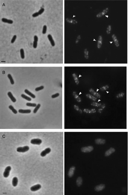
Fig. 1.
Localization of MurG in wild-type E. coli LMC500 (A) and in the MurG(Ts) strain GS58 (B and C). Phase-contrast images are given on the left and fluorescence images on the right. LMC500 cells were grown to steady state at 28°C in GB1 (A), fixed, permeabilized and immunolabelled with anti-MurG. GS58 cells were first grown to mid-exponential phase at 28°C in TY medium (B) shifted to 42°C and allowed to grow for 2 MDs (C). Thereafter they were fixed, permeabilized and subjected to MurG IFM. The arrows point to the band at mid-cell. All panels have the same exposure time. Scale bar equals 1 μm.

