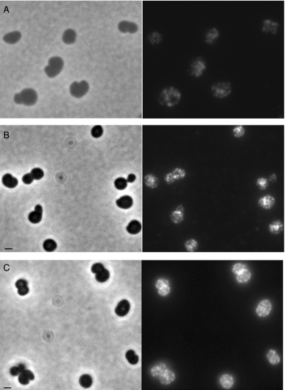
Fig. 7.
MurG and MreB localization are independent of the spherical cell morphology (A and B). LMC500 cells were grown for 2 MDs in the presence of mecillinam (inhibitor of PBP2), at 28°C in GB1. Cells were immunolabelled with anti-MreB (A) or anti-MurG (B). The MreB helical structure is preserved and the multi-foci localization pattern of MurG is observed. Arrangement of MurG is dependent on the presence of MreCD (C). IFM was performed with PA340-678pMEW1 strain (mreBCD deletion strain with the pMEW1 plasmid that expresses MreC and MreD constitutively) grown at 28°C in TY to mid-exponential phase. In these spherical cells MurG localized normally as multiple foci in the cell envelope and at mid-cell. Phase contrast (left) and fluorescence images (right) are shown. Scale bar equals 1 μm.

